


RESEARCH AREAS
Bioingénierie Tissulaire (BioTis)
Inserm U1026
Université Bordeaux
146, rue Léo-Saignat
33076 BORDEAUX cedex - France
Tél. : 33 (0)5 57 57 14 88
Fax : 33 (0)5 56 90 05 17
Cardiovascular applications have been at the center of BioTis's research activity for over 20 years. This unit pioneered the culture of human endothelial cells from different sources including cord blood adult stem cells and has been at the forefront of cardiovascular tissue-engineering in France. Current projects are focused on the development of small diameter (≤ 5 mm) vascular grafts and on the production of vascular networks.
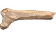
Functional small diameter vascular grafts are needed for heart bypass, leg bypass and brain surgery. However, the current synthetic grafts perform very poorly in these indications and, as a result, they are only acceptable for leg revascularization procedures. Two main approaches are under investigation. First, the use of a chitosan-based scaffold to produce a cell-free hemocompatible vascular graft that can also support endothelial cell colonization. This project is led by Pr. Laurence Bordenave in collaboration with the “Laboratoire d’Ingénierie des Matériaux Polymères (IMP)” (UMR 5223, Villeurbanne, Pr. Laurent DAVID) Recent data has shown promising preliminary results in vivo in a large animal model. This project was funded by the “Agence Nationale de Recherche (ANR)” (TecSan program).
Chitosan-based vascular grafts under development at BioTis.
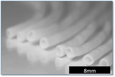
A second approach under development at BioTis is the use of Cell-Assembled extracellular Matrix (CAM) produced by normal human fibroblasts in vitro.[2,3] With the arrival of Nicolas L'Heureux, this technology, which was first developed in his company in California, will be introduced in Europe and will be used for the development of a new generation of small diameter vascular grafts. These new grafts will be built from yarn made of CAM and assembled with a textile-inspired method. As a result, the grafts will be produced 3 times faster than previously and will have better and more tunable mechanical properties. This is a unique approach that can also produce human textiles for various applications.
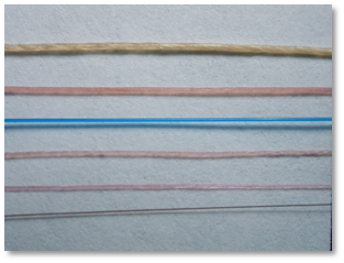
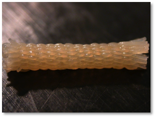
Threads of various sizes, 5-0 Prolene (bleu) and human hair (bottom). On the right, a woven TEBV prototype of 2.2 mm I.D,
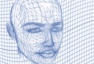
The third vascular project is headed by Pr. Raphaël Devillard and aims at the development of vascular networks. These networks are a critical component of any bulk tissue and represent one of the key challenges in tissue engineering for the next decades. Indeed, for the development of pancreas, kidney, liver, lung, brain, etc., the ability to create a graftable hierarchically organized vascular tree is critical. At BioTis, vascular networks are of particular interest because they have emerged as key players in bone hemostasis and repair (see the Bone Engineering section). As a result, this project is at the interface of the core capabilities of BioTis: vascular tissue engineering, bone tissue engineering and biofabrication. Indeed, BioTis is a leader in bioprinting in France[4,5](see the Biofabrication section) and has recently spun off a startup (Poietis). Strategies to produce vascular networks include printing endothelial (± other cells) in complex patterns and on various substrates including biodegradable polymers and biological matrices such as the CAM and the amniotic membrane.
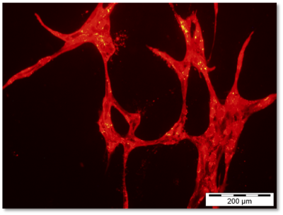
Human endothelial cells printed to form a vascular network
1. Rami, L., et al. Physicochemical modulation of chitosan-based hydrogels induces different biological responses: interest for tissue engineering. J. Biomed. Mater. Res. A 102, 3666-3676 (2014).
2. Peck, M., Dusserre, N., McAllister, T.N. & L'Heureux, N. Tissue engineering by self-assembly. Materials Today 14, 218-224 (2011)
3. Peck, M., Gebhart, D., Dusserre, N., McAllister, T.N. & L'Heureux, N. The evolution of vascular tissue engineering and current state of the art. Cells Tissues Organs 195, 144-158 (2012).
4. Devillard, R., et al. Cell patterning by laser-assisted bioprinting. Methods Cell Biol. 119, 159-174 (2014).
5. Ali, M., Pages, E., Ducom, A., Fontaine, A. & Guillemot, F. Controlling laser-induced jet formation for bioprinting mesenchymal stem cells with high viability and high resolution. Biofabrication 6, 045001 (2014).